


Varicose veins of the lower extremities are characterized by dilation of the superficial veins of the legs, accompanied by the destruction of the blood flow of the legs and the failure of the valves. As a result, the length and diameter of the veins increase, giving a serpentine, cylindrical, or saccular appearance, although there are also mixed manifestations of the listed deformities.
Characteristics of the venous system
The appearance and development of varicose veins are directly related to the venous system of the legs, including:
- Great saphenous vein: small and large;
- Deep veins (in the calves and thighs);
- Perforating vein, this is the link between the first two systems.
Under normal circumstances, 90% of the blood is delivered to the lower extremities through the deep veins, and the remaining 10% is delivered to the lower extremities through the superficial veins. When it returns to the side of the heart, this mechanism is supported by valves in the vein wall. When the next part of blood arrives, they will violently hit to prevent it from moving from top to bottom under the influence of gravity. Muscle contraction pushes blood further to the heart, allowing blood to flow normally.
A person who stays upright for a long time may cause blood to stagnate, which increases the pressure of the vein and causes its diameter to increase. This process can cause incomplete closure of the valve leaflets, and as a result, the blood flow is disturbed by the backflow from the heart-backflow.
Deep venous valves are most likely to be affected because they carry the largest amount of blood and therefore bear the greatest load. In order to reduce their high pressure, part of the blood is delivered to the superficial surface through the perforated vein, which was not originally intended for mass use. This load on the vein walls causes their expansion and the formation of varicose veins.
At the same time, blood continuously enters the deep veins, but due to the destruction of the function of the deep veins and the normal activity of the perforated vein valve leaflets, the blood is redistributed to the superficial vessels. As a result, chronic varicose veins develop, accompanied by pain, edema, and trophic ulcers over time.
Cause
In the past, one of the main causes of varicose veins was called genetic factors, but today this theory has been refuted. Of course, it is possible to track the frequent manifestations of the disease in some families, but this is more likely to be due to the life characteristics taught in the family: dietary culture, passive rest, sedentary, etc.
The development of varicose veins is based on the presence of reflux in the venous system, when blood circulates through the veins in the opposite direction. Due to congenital or acquired degeneration of the valve device, additional transport of blood from deep veins to superficial veins is possible. When venous knots form, this can cause superficial blood vessels to overfill and dilate.
One of the root causes of the development of varicose veins is believed to be an unhealthy diet, which in some cases can lead to obesity. Such people do not move much, mainly eat highly processed foods, and minimize the proportion of plant fiber in their diet. After all, it is they are involved in strengthening veins and blood vessel walls and preventing long-term chronic constipation, which can greatly increase intra-abdominal pressure and cause varicose veins. It is worth noting that a weight gain of more than 20% will increase the risk of disease five-fold.
The main cause of women is childbirth, and each pregnancy increases the risk of varicose veins. Severe weight gain and enlarged uterus put a lot of pressure on the legs, causing the legs to stagnate. This situation is exacerbated by the increasing intra-abdominal pressure and the action of progesterone hormone, which affects the state of the elastic fibers of the blood vessel wall.
Other factors that cause varicose veins in the lower extremities include:
- A sedentary lifestyle, standing upright during the day (for example, a hairdresser), flying long distances or traveling long distances. All of these can cause stagnation in the lower limbs, when blood accumulates in the superficial veins and is difficult to transport to the heart;
- Sometimes it increases the risk of women suffering from varicose veins, wearing uncomfortable tight shoes, especially models with high heels;
- Corsets and tight underwear squeeze the groin veins and increase intra-abdominal pressure, which is the direct prerequisite for varicose veins;
- hypertension;
- Smoking, which indirectly leads to thinning of blood vessel walls.
Disease classification
The classification of varicose veins of the lower extremities depends on the incidence of venous disease, their location and the existence of pathological reflux, which is characterized by impaired blood outflow. There are 4 forms of varicose veins:
- Intradermal and subcutaneous varicose veins (segmental), in which there is no pathological outflow of venous blood;
- Segmental varicose veins, when reflux occurs through perforating veins or superficial veins;
- A common form of varicose veins in which reflux occurs through both the perforating and superficial veins;
- Varicose veins are characterized by deep venous return.
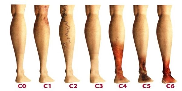
After the chronicity of varicose veins of the lower extremities, phlebology believes that it has three degrees:
- Temporary edema occurs periodically in the context of "heavy legs" syndrome.
- Persistent and persistent edema. Hyperpigmentation and eczema may occur.
- Nutritional venous ulcer.
The latter is the most difficult to treat because it requires initial removal of inflammation and healing of skin tissue.
Stages and symptoms
The disease progresses very slowly, sometimes lasting for more than ten years, until the onset of symptoms force the patient to seek advice from a phlebologist. In the early stages of varicose veins, their manifestations are often attributed to fatigue, age or other reasons. In order to fully consider the symptoms of the disease, the manifestations of varicose veins are classified according to the stage:
- The first stage occurs more often when you are young-20 years later, when there is a feeling of heaviness in the legs, edema may appear and disappear completely at night. On the inner side of the calf, you can see enlarged veins that appear as lumps of skin. At this stage, many people noticed small spider veins. In general, symptoms are subtle and rarely receive the attention they deserve.
- The second stage is characterized by increased external manifestations of dilated veins. In the context of the pathological work of venous valves, the disease has progressed, therefore, the size of the saphenous veins has increased significantly, and their elongation can also be noticed. It is more common that there is a feeling of heaviness and burning in the legs, and they will soon feel tired from the long journey.
- The disease has become a chronic disease due to the continuous imbalance of venous blood flow. At night, the patient will develop near-ankle edema, which can be very serious. Heaviness in the legs and cramps at night.
- Without treatment in the previous stage, chronic insufficiency of the function of the venous system will negatively affect the metabolism of the skin, especially the lower leg area. Darkening of the skin can be seen near the ankle-hyperpigmentation, it will thicken and become inflamed over time. The condition described is called steatodermal sclerosis. If you do not start to treat the venous system at this time, trophic ulcers will soon begin to form.
- The fifth stage is accompanied by many trophic ulcers, some of which heal periodically as scars form.
- Extensive ulcers will appear in areas with long-standing nutritional disorders. This condition requires urgent and active treatment, aimed at the healing of varicose veins and skin ulcers.
diagnosis
Perform external examination of the lower limbs in the vertical and horizontal positions of the body, venous palpation, and preliminary assessment of the stage of the disease. The patient is sent for a general blood test, which allows you to study the picture of the disease more thoroughly:
- At the platelet level, it will reflect the tendency of thrombosis;
- The level of hemoglobin and the number of red blood cells indicate the degree of blood clotting;
- By increasing the level of white blood cells, you can determine whether there is inflammation, which helps to diagnose thrombophlebitis more quickly.
Be sure to check the venous system of the legs. There are many ways:
- Ultrasound Doppler imaging-USDG;
- Venography;
- CT venography;
- Duplex vascular scan-USAS;
- Venography;
- Photoplethysmography;
- Venous manometry and so on.
In practice, patients use USAS and USG more often because they help to comprehensively study the venous system of the legs and identify degenerative areas. If the ultrasound examination does not give a complete view of the disease image, the remaining methods can be additionally specified. Some of these methods may have complications, such as venous thrombosis, catheter perforation, and hypersensitivity to contrast agents. Consider the most commonly used techniques in phlebology:
- USAS allows assessment of the anatomical, hemodynamic and functional pathology of the venous bed. The data obtained is processed by the computer, and then the model of the venous system can be viewed on video or printed on paper.
- High-accuracy Doppler ultrasonography determines the patency and blood flow velocity of superficial and deep veins. Doppler ultrasound can evaluate the function of the valve device.
After extensive diagnosis, the doctor will draw the patient's vein card, which allows you to determine the damaged part, extent and length of the venous system. After that, choose the right treatment.
treatment
It is carried out in a comprehensive manner and is determined according to the symptoms, the degree of disease development, and the results of research. In the initial stage, conservative treatment is prescribed, which includes:
- Medication when prescribing a group of medications:
- Anti-protective agents and intravenous injections;
- Anticoagulant
- Decomposition
- Topical preparations (ointments, gels);
- Anti-inflammatory drugs.
- For elastic compression, use compression stockings or bandages (rarely). It allows you to dose the squeeze of the muscles, prevent stagnant processes, and improve blood flow through the blood vessels. When wearing this kind of underwear, it has the effect of artificially maintaining blood vessel tension.
- Physical therapy methods, the best results of which are electrophoresis, radial current, laser radiation and magnetic fields.
- Viable physical activity can only be done in compression underwear (except for swimming). It is recommended to ride a bicycle, swim, and jog. The phlebologist chooses a set of individual exercises for the lower limbs and trains the blood vessels in the legs every day.
In addition, it is recommended that patients perform a five-minute comparison program in the shower every night, alternately switching from warm water to cold water. Such operations can improve blood flow and regulate blood vessels.
It is important to determine the factors that induce the disease at the beginning of treatment to effectively influence it. At-risk patients should see a phlebologist every 2 years for preventive examinations and ultrasound examinations of leg veins.
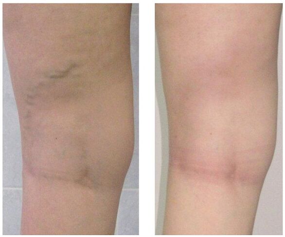
When conservative treatment has no results or when varicose veins are observed in the late stage, surgical intervention is used. Today, varicose veins can be completely cured by:
- Phlebectomy. The essence of the operation is to remove the main superficial vein to eliminate pathological blood discharge. The perforating vein is often ligated to achieve the same purpose.
- Sclerotherapy. It involves introducing sclerosing agent into the affected area of the vein, thereby connecting its walls. Recently, they have begun to actively use foam hardeners for the same purpose based on technology. The blood flow through the defect area is stopped, and cosmetic defects in the form of prominent nodules are eliminated. After such intervention, no scars were left. All operations were performed in outpatient clinics, and there was no follow-up hospitalization. But sclerotherapy is only used for the fusion of small branches of the vein trunk.
- Laser coagulation. With the help of a laser beam, the marked part of the vein is heated, the vein walls stick together, and blood flow stops. But this technique is only suitable for dilating veins less than one centimeter in diameter.
prevention
Preventive measures can be primary, aimed at preventing the development of varicose veins, or secondary, when it is necessary to reduce the risk of recurrence after surgery or prevent the course of the disease from getting worse. Helpful hints:
- Live an active lifestyle that does not put a heavy burden on your legs: swimming, walking, biking;
- Pay attention to your weight;
- Lift your legs more frequently;
- Do not wear tight underwear and high heels of 4 cm or more;
- Use orthopedic insoles;
- Take a contrast shower;
- Do five minutes of preventive leg exercises every day;
- Wear stretch stockings for long walks.
If you find the slightest varicose veins-prominent nodules, swelling, and heaviness in the legs, please do not postpone your visit to a phlebologist. In fact, over time, this hidden disease can cause many complications, including thrombophlebitis and thrombosis.