


A disease caused by the destruction of the structure of the blood vessel wall, the thinning of the blood vessel wall and the obvious stagnation of blood flow, is called varicose veins. This disease usually affects the lower extremities, although it can be localized to other parts of the body. According to the tenth revised International Classification of Diseases in ICD 10, varicose veins are designated as code 183, which includes four headings describing various manifestations of the disease.
How do varicose veins appear?
The first mention of varicose veins was found in ancient Greek papyrus. History and proven scientific facts indicate that varicose veins of the lower limbs were found in Egyptian mummies—it can be said that this disease is accompanied by the entire existence of human beings.
Outstanding doctors-Avicenna, Hippocrates, Galen tried to treat varicose veins of the lower limbs. In the 19th century, painful and traumatic treatments were used, including dissecting the tissues of the thigh and calf to damage the saphenous vein and then bandaging it. It is understood that in this way the stagnant blood flow process can be prevented and varicose veins can be eliminated. However, these methods left terrible, extensive scars on patients, and resulted in nerve and arterial damage and interruption of lymphatic flow.
Soon after, the history of the treatment of varicose veins made a positive breakthrough-in 1908, the metal probe was used for the first time as a means of micro-creation of the blood vessel wall. The discovery of radiography has made it possible to perform more accurate and effective surgical procedures to eliminate varicose veins. Now, when it is necessary to correctly diagnose and treat diseases, double and triple scanning, powerful drugs, laser therapy and sclerotherapy are used. Surgical intervention is only used when varicose veins cannot be eliminated in a controlled manner.
The main cause of the disease
Varicose veins are very harmful, and the pathology has become "younger". In the past, most people suffering from varicose veins were elderly people. Now varicose veins are more common in young patients, and very rare in children.
Causes:
- Genetic susceptibility.
- Overweight, overweight, obese.
- Sedentary lifestyle.
- Improper diet and poor blood quality.
- Concomitant diseases of the cardiovascular system.
- Professional activities.
- Standing for a long time, heavy physical exertion.
- Pregnancy and hormonal changes.
- Individual characteristics of the structure of the vascular system.
- Pathological congenital disease.
- Wear high-heeled shoes and tight-fitting clothes.
- Heat treatment.
Any of the above causes may cause the development of varicose veins, and the consequences are dangerous, including death.
The structure of the veins
To understand the causes of varicose veins in the lower extremities, you need to understand the structure of the vascular system and its working mechanism. It represents the sum of the main (deep and superficial) and connecting (connecting) veins.
The superficial superficial venules start from the foot and extend along the back of the calf. There are two branches under the knee that connect with the popliteal vein and the deep femoral vein.
A large superficial saphenous vein is formed in the ankle area, extending along the surface of the calf and knee joint, and connected to the femoral vein. The deep veins are distributed along the branches of the arteries, and the entire venous system is connected by perforating blood vessels.
When blood flow is normal, oxygenated blood flows directly to the heart, and special venous valves can prevent backflow. Varicose veins of the lower extremities mean strong pressure, the diameter of the venous lumen increases significantly, the valve cannot cope with the task, and a backflow-reverse blood flow occurs. Abnormal blood circulation can cause excessive expansion (stretching) of blood vessel walls, thinning, venous obstruction and blood stasis. Result-abdominal distension, swelling of veins, formation of nodes.
Symptoms and clinical manifestations
Varicose veins can progress latently for a long time, and then the following signs appear:
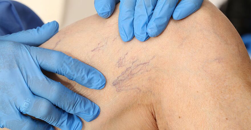
- The formation of spider veins is a web-like accumulation of dilated venules.
- A clear pattern of hyperemic veins protruding under the skin.
- The formation of vascular compaction-varicose veins in the legs, forming clearly discernible nodules.
- The normal color of the skin changes, cyanosis appears, darkens, and the upper layer (dermis) acquires a loose structure.
- Feeling of pain, heaviness, swelling and fatigue in the legs, inconvenience in movement, and difficulty in walking.
- With varicose veins of the lower extremities, the formation of soft tissue swelling is possible.
Neglecting timely treatment can lead to serious and dangerous consequences, and only through immediate surgical intervention can a person be cured.
Disease classification
According to ICD 10, varicose veins are classified as ulcer disease, inflammatory disease, ulcer disease and inflammation when these symptoms are not present. According to the International Classification of Chronic Venous Diseases established in 1994, varicose veins are divided into:
- Intradermal, segmental. No pathological venous secretions were observed.
- Segments with reverse blood flow occur through superficial veins and perforating veins.
- Distribution and reverse blood flow through superficial veins and perforating veins.
- With varicose veins, blood flows back through deep veins.
Diseases are usually classified according to other signs of clinical manifestations:
- On examination or palpation, there were no symptoms.
- Reticulate vein expression.
- There are varicose veins.
- There is swelling of soft tissues.
- Violation of normal skin tone.
- Found fatty skin sclerosis.
- There is a healed ulcer.
- An active ulcer was found.
Symptoms are absent or subjective (what the patient feels). In addition, the reasons for the classification of varicose veins are: congenital, primary, secondary, and unknown factors that lead to the development of the disease.
Diagnosis of varicose veins
The main method of identifying varicose veins is to visually inspect and palpate the patient. In order to carefully determine the severity of the disease and choose the correct treatment method, when studying the medical history and performing palpation, the phlebologist prescribes the following:
- A complete blood count is the main criterion for determining the number of red blood cells and the level of hemoglobin. Based on blood coagulation, conclusions are drawn about the degree of disease progression and the tendency to thrombosis.
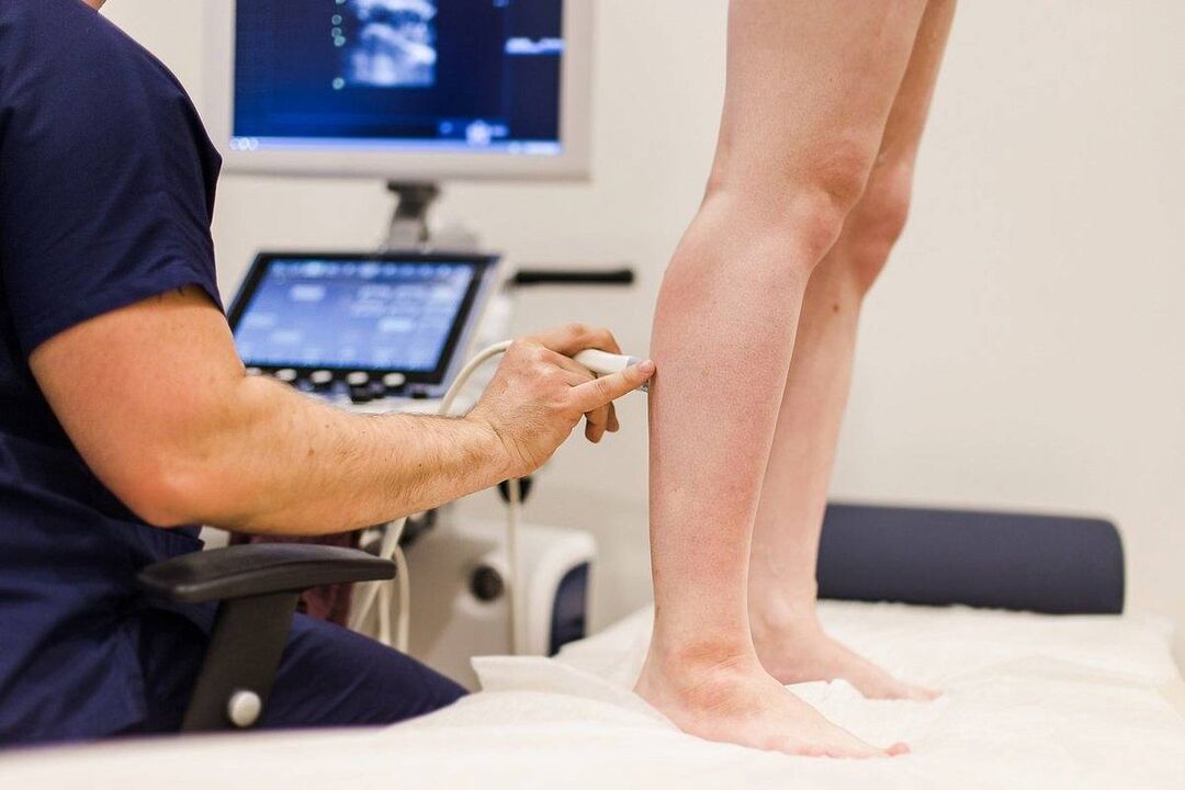
- Doppler ultrasound. The method includes ultrasound diagnosis of the velocity and direction of blood particles. This allows you to determine the direction of blood flow and whether there is sufficient speed.
- Ultrasound agnioscanning. It is used to visually inspect the blood vessel wall and its structure, blood flow direction and speed in real time on the monitor of the ultrasound machine.
- Plethysmography. The diagnosis is based on detecting the electrical resistance of the leg tissue. In the appropriate cycle, this parameter should indicate the normal standard.
- Rheological diagnosis. Based on the determination of tissue blood filling index. The rheological index helps determine the stage of varicose veins-compensatory, off-compensation, or decompensation.
Medical history and research, obtaining comprehensive diagnostic data, allowing doctors to choose treatment methods.
Conservative medication
This treatment includes the use of special drugs that have a positive effect on the course of the disease. The conservative treatment of varicose veins is effective at the initial stage and is used as an additional method to treat lymph nodes, ulcers, and eczema.
The main drug groups prescribed are:
- Intravenous injection and intravenous protective agent. Venotonic drugs are standard, meaning conservative treatment. Promote the repair of blood vessel wall structure, strengthen and regulate blood vessels.
- Effective way to thin blood. They help improve quality components, make blood flow faster in veins, reduce the risk of blood clots, restore normal blood circulation, and relieve pain.
- NSAID (anti-inflammatory drug). Eliminate pain, prevent cramps, and effectively relieve inflammation and swelling.
Conservative treatment is helpful for timely referral to a phlebologist, which may affect the blood composition and the condition of the blood vessel wall in the initial stage. For the complex forms of the disease, severe measures need to be taken.
Laser Treatment
When varicose veins in the legs need to be classified according to ICD 10 code 183, laser treatment is considered a gentle and minimally invasive method. The idea of this method is to use a laser beam to positively affect the blood vessel wall and promote its adhesion. The LED connected to the laser device is inserted into the vein by piercing the skin. The beam is selective and has no effect on adjacent healthy tissues. Significant advantages of laser treatment of varicose veins:
- Fast positive effect.
- There is no pain or injury.
- The result is stable and long-term relief.
- Restore normal blood circulation.
Contraindications for use are thick or thin blood vessel walls, large venous cavities, pregnancy, tumors and other serious concomitant diseases.
Sclerotherapy for varicose veins
The method is based on introducing a special liquid or foam formulation-a sclerosant into the blood vessels affected by varicose veins. They use fibrous tissue instead of endothelial cells. To perform sclerotherapy, needles, syringes, and sclerosing agents are used.
The processing technique includes the following steps:
- Puncture pathological veins;
- Draw (remove) all blood from the blood vessel;
- Administration of hardener preparations;
- Apply appropriate bandages or knitted compresses.
This method gives lasting results. The procedure is painless, and the fusion of vascular tissue and varicose veins is an alternative to surgery.
Perform operation
The most painful and traumatic way to treat varicose veins is surgery. The indications for execution are extensive vascular disease, the presence of varicose veins, and the dangerous consequences of the disease, such as acute thrombophlebitis.
Phlebectomy is performed under local anesthesia. The pathological vein is ligated, the required number of incisions are made to remove it, and then it is removed. This operation is recognized as an effective treatment method, showing results in 80% of cases. But phlebectomy has many side effects: wound complications, lymph node trauma, and in extreme cases, it can cause deep nerve damage, restricted mobility and disability.
In order to prevent the dangerous complications of varicose veins, which are manifested as: nodules, ulcers, bleeding, venous thrombosis, pulmonary embolism and other serious consequences, consult a doctor in the early stage of varicose veins!